Confocal microscopy — Diagnostics MeSH D018613 OPS 301 code 3 301 … Wikipedia
confocal microscopy — A system of (usually) epifluorescence light microscopy in which a fine laser beam of light is scanned over the object through the objective lens. The technique is particularly good at rejecting light from outside the plane of focus, and so… … Dictionary of molecular biology
confocal microscopy — noun An imaging technique in which pinholes are used to eliminate out of focus light … Wiktionary
Confocal laser scanning microscopy — (CLSM or LSCM) is a technique for obtaining high resolution optical images with depth selectivity.[1] The key feature of confocal microscopy is its ability to acquire in focus images from selected depths, a process known as optical sectioning.… … Wikipedia
Microscopy — is the technical field of using microscopes to view samples and objects that cannot be seen with the unaided eye (objects that are not within the resolution range of the normal eye). There are three well known branches of microscopy, optical,… … Wikipedia
Confocal — In geometry, confocal means having the same foci. For an optical cavity consisting of two mirrors, confocal means that they share their foci. If they are identical mirrors, their radius of curvature, Rmirror, equals L, where L is the distance… … Wikipedia
Scanning confocal electron microscopy — (SCEM) is an electron analogue of scanning confocal optical microscopy (SCOM) where electrons and electron lenses are used instead of light and optical lenses. Advantages of SCEM High energies of incident particles (200 keV electrons vs. 2 eV… … Wikipedia
Two-photon excitation microscopy — is a fluorescence imaging technique that allows imaging of living tissue up to a very high depth, that is up to about one millimeter. Being a special variant of the multiphoton fluorescence microscope, it uses red shifted excitation light which… … Wikipedia
Microscopio confocal — Principios en los que se basa la microscopía confocal. El microscopio confocal es un microscopio que emplea una técnica óptica de imagen para incrementar el contraste y/o reconstruir imágenes tridimensionales utilizando un pinhole espacial… … Wikipedia Español
Microscope confocal — Pour les articles homonymes, voir Microscope (homonymie) et Microscopie. Schéma de principe du microscope confocal par Marvin Minsky en 1957 Un micr … Wikipédia en Français

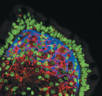
 Confocal microscopy used to examine fluorescent in situ hybridization of a small intestinal biopsy specimen in a patient with Whipple disease. The intestinal villus shows ribosomal RNA (rRNA) of the infecting bacteria Tropheryma whipplei (blue), as well as human cell nuclei (green), and vimentin (red).
